Sample Work
Editing Samples
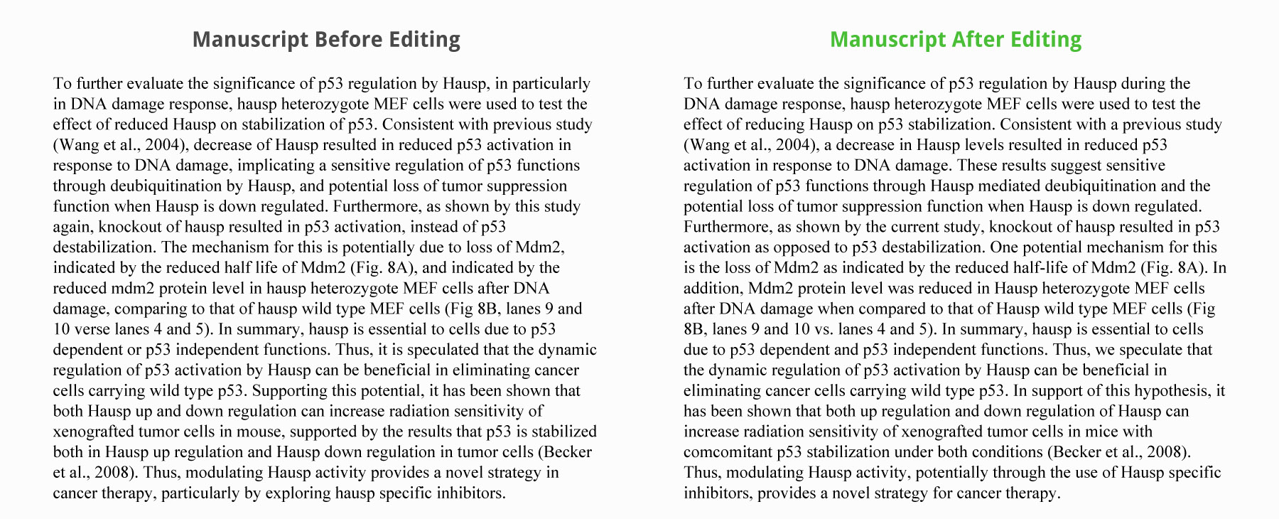
Journal Submission: Nature Publication Group
The client was submitting their manuscript as a new submission to a Nature Publication Group journal and needed assistance with improving on the language. They also needed help reducing the word count of the abstract.
Our Editors:
- removed all spelling and grammar mistakes
- shortened redundant sentences
- made sentences more succinct and direct
- offered formatting recommendations
- suggested ways to re-word sentences reduce the word count without changing the meaning
Sample (click to open):
Manuscript Before Editing
To further evaluate the significance of p53 regulation by Hausp, in particularly in DNA damage response, hausp heterozygote MEF cells were used to test the effect of reduced Hausp on stabilization of p53. Consistent with previous study (Wang et al., 2004), decrease of Hausp resulted in reduced p53 activation in response to DNA damage, implicating a sensitive regulation of p53 functions through deubiquitination by Hausp, and potential loss of tumor suppression function when Hausp is down regulated. Furthermore, as shown by this study again, knockout of hausp resulted in p53 activation, instead of p53 destabilization. The mechanism for this is potentially due to loss of Mdm2, indicated by the reduced half life of Mdm2 (Fig. 8A), and indicated by the reduced mdm2 protein level in hausp heterozygote MEF cells after DNA damage, comparing to that of hausp wild type MEF cells (Fig 8B, lanes 9 and 10 verse lanes 4 and 5). In summary, hausp is essential to cells due to p53 dependent or p53 independent functions. Thus, it is speculated that the dynamic regulation of p53 activation by Hausp can be beneficial in eliminating cancer cells carrying wild type p53. Supporting this potential, it has been shown that both Hausp up and down regulation can increase radiation sensitivity of xenografted tumor cells in mouse, supported by the results that p53 is stabilized both in Hausp up regulation and Hausp down regulation in tumor cells (Becker et al., 2008). Thus, modulating Hausp activity provides a novel strategy in cancer therapy, particularly by exploring hausp specific inhibitors.
Edited Manuscript
To further evaluate the significance of p53 regulation by Hausp during the DNA damage response, hausp heterozygote MEF cells were used to test the effect of reducing Hausp on p53 stabilization. Consistent with a previous study (Wang et al., 2004), a decrease in Hausp levels resulted in reduced p53 activation in response to DNA damage. These results suggest sensitive regulation of p53 functions through Hausp mediated deubiquitination and the potential loss of tumor suppression function when Hausp is down regulated. Furthermore, as shown by the current study, knockout of hausp resulted in p53 activation as opposed to p53 destabilization. One potential mechanism for this is the loss of Mdm2 as indicated by the reduced half-life of Mdm2 (Fig. 8A). In addition, Mdm2 protein level was reduced in Hausp heterozygote MEF cells after DNA damage when compared to that of Hausp wild type MEF cells (Fig 8B, lanes 9 and 10 vs. lanes 4 and 5). In summary, hausp is essential to cells due to p53 dependent and p53 independent functions. Thus, we speculate that the dynamic regulation of p53 activation by Hausp can be beneficial in eliminating cancer cells carrying wild type p53. In support of this hypothesis, it has been shown that both up regulation and down regulation of Hausp can increase radiation sensitivity of xenografted tumor cells in mice with comcomitant p53 stabilization under both conditions (Becker et al., 2008). Thus, modulating Hausp activity, potentially through the use of Hausp specific inhibitors, provides a novel strategy for cancer therapy.
With Track Changes
Accepted and Published!
The manuscript was sent for review and received positive critiques. We re-edited the manuscript after the revisions were complete for free, since all re-edits on the same manuscript are included in our low fee. The paper was accepted and published upon re-submission.
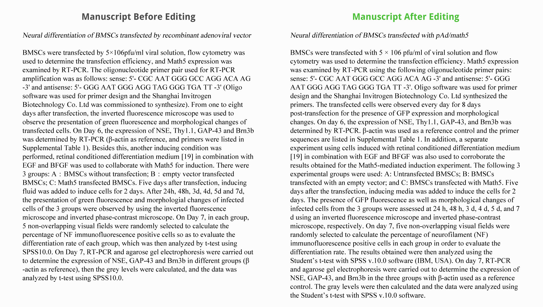
Journal Submission: Cell
The client had submitted their manuscript to a Cell journal and received positive critiques from the reviewers. However, one of the criticisms was poor language usage. The client contacted us to help improve the language for re-submission.
Our Editors:
- improved the grammar
- suggested removing superfluous descriptions
- helped add clarity to the discussion section
- offered formatting suggestions of the tables
Sample (click to open):
Manuscript Before Editing
Neural differentiation of BMSCs transfected by recombinant adenoviral vector
BMSCs were transfected by 5×10⁶pfu/ml viral solution, flow cytometry was used to determine the transfection efficiency, and Math5 expression was examined by RT-PCR. The oligonucleotide primer pair used for RT-PCR amplification was as follows: sense: 5'- CGC AAT GGG GCC AGG ACA AG -3' and antisense: 5'- GGG AAT GGG AGG TAG GGG TGA TT -3' (Oligo software was used for primer design and the Shanghai Invitrogen Biotechnology Co. Ltd was commissioned to synthesize). From one to eight days after transfection, the inverted fluorescence microscope was used to observe the presentation of green fluorescence and morphological changes of transfected cells. On Day 6, the expression of NSE, Thy1.1, GAP-43 and Brn3b was determined by RT-PCR (β-actin as reference, and primers were listed in Supplemental Table 1). Besides this, another inducing condition was performed, retinal conditioned differentiation medium [19] in combination with EGF and BFGF was used to collaborate with Math5 for induction. There were 3 groups: A:BMSCs without transfection; B:empty vector transfected BMSCs; C: Math5 transfected BMSCs. Five days after transfection, inducing fluid was added to induce cells for 2 days. After 24h, 48h, 3d, 4d, 5d and 7d, the presentation of green fluorescence and morphologial changes of infected cells of the 3 groups were observed by using the inverted fluorescence microscope and inverted phase-contrast microscope. On Day 7, in each group, 5 non-overlapping visual fields were randomly selected to calculate the percentage of NF immunofluorescence positive cells so as to evaluate the differentiation rate of each group, which was then analyzed by t-test using SPSS10.0. On Day 7, RT-PCR and agarose gel electrophoresis were carried out to determine the expression of NSE, GAP-43 and Brn3b in different groups (β-actin as reference), then the grey levels were calculated, and the data was analyzed by t-test using SPSS10.0.
Edited Manuscript
Neural differentiation of BMSCs transfected with pAd/math5
BMSCs were transfected with 5 × 10⁶ pfu/ml of viral solution and flow cytometry was used to determine the transfection efficiency. Math5 expression was examined by RT-PCR using the following oligonucleotide primer pairs: sense: 5'- CGC AAT GGG GCC AGG ACA AG -3' and antisense: 5'- GGG AAT GGG AGG TAG GGG TGA TT -3'. Oligo software was used for primer design and the Shanghai Invitrogen Biotechnology Co. Ltd synthesized the primers. The transfected cells were observed every day for 8 days post-transfection for the presence of GFP expression and morphological changes. On day 6, the expression of NSE, Thy1.1, GAP-43, and Brn3b was determined by RT-PCR. β-actin was used as a reference control and the primer sequences are listed in Supplemental Table 1. In addition, a separate experiment using cells induced with retinal conditioned differentiation medium [19] in combination with EGF and BFGF was also used to corroborate the results obtained for the Math5-mediated induction experiment. The following 3 experimental groups were used: A: Untransfected BMSCs; B: BMSCs transfected with an empty vector; and C: BMSCs transfected with Math5. Five days after the transfection, inducing media was added to induce the cells for 2 days. The presence of GFP fluorescence as well as morphological changes of infected cells from the 3 groups were assessed at 24 h, 48 h, 3 d, 4 d, 5 d, and 7 d using an inverted fluorescence microscope and inverted phase-contrast microscope, respectively. On day 7, five non-overlapping visual fields were randomly selected to calculate the percentage of neurofilament (NF) immunofluorescence positive cells in each group in order to evaluate the differentiation rate. The results obtained were then analyzed using the Student’s t-test with SPSS v.10.0 software (IBM, USA). On day 7, RT-PCR and agarose gel electrophoresis were carried out to determine the expression of NSE, GAP-43, and Brn3b in the three groups with β-actin used as a reference control. The gray levels were then calculated and the data were analyzed using the Student’s t-test with SPSS v.10.0 software.
With Track Changes
Accepted for Publication!
The manuscript was re-submitted and accepted by the journal.
Translation Samples
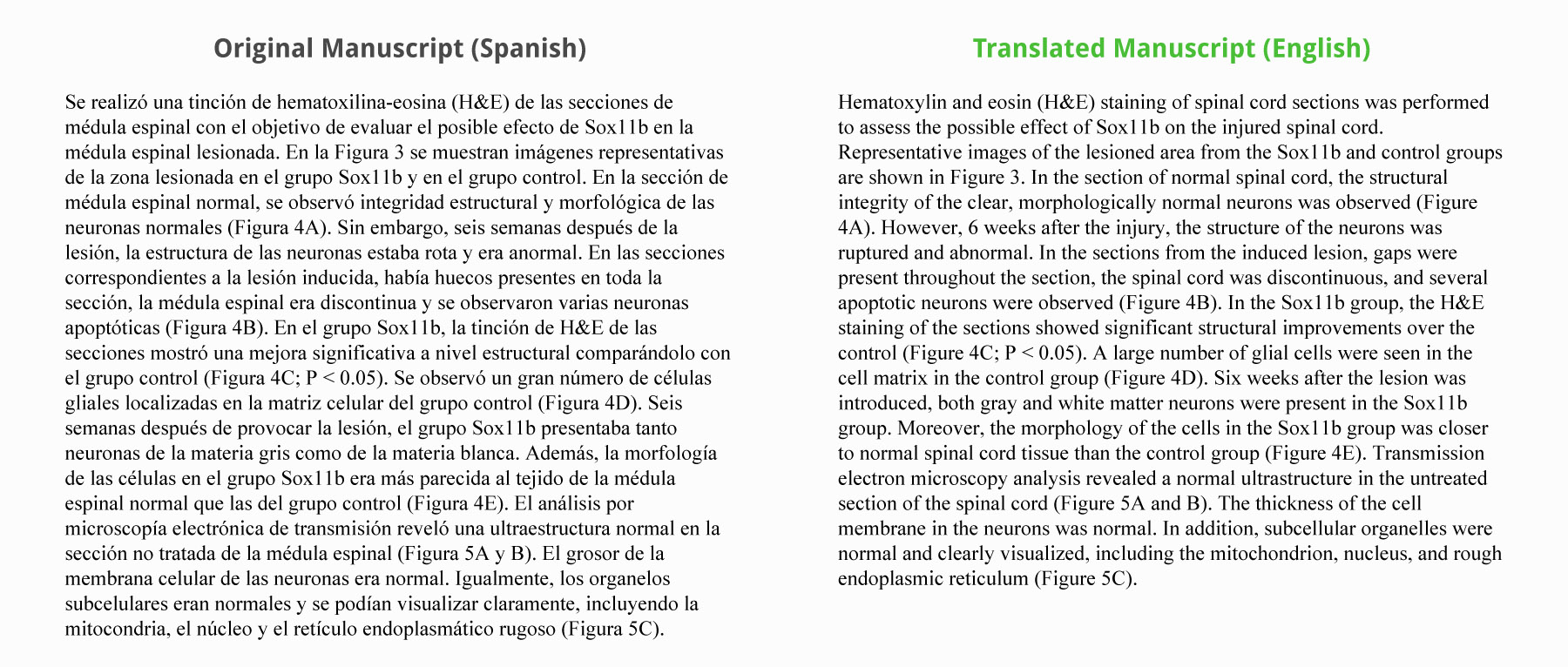
Journal Submission: ASBMB journal
The client communicated with our Spanish bilingual editor and asked for a complete translation of their manuscript to English. A native English speaking editor then completed a second review of the document.
Our Editors:
- translated the manuscript from Spanish to English
- a native English speaking editor performed the secondary edits
- confirmed the correct scientific translation with both the author and editors
Sample (click to open):
Original Scientific Manuscript in Spanish
Se realizó una tinción de hematoxilina-eosina (H&E) de las secciones de médula espinal con el objetivo de evaluar el posible efecto de Sox11b en la médula espinal lesionada. En la Figura 3 se muestran imágenes representativas de la zona lesionada en el grupo Sox11b y en el grupo control. En la sección de médula espinal normal, se observó integridad estructural y morfológica de las neuronas normales (Figura 4A). Sin embargo, seis semanas después de la lesión, la estructura de las neuronas estaba rota y era anormal. En las secciones correspondientes a la lesión inducida, había huecos presentes en toda la sección, la médula espinal era discontinua y se observaron varias neuronas apoptóticas (Figura 4B). En el grupo Sox11b, la tinción de H&E de las secciones mostró una mejora significativa a nivel estructural comparándolo con el grupo control (Figura 4C; P < 0.05). Se observó un gran número de células gliales localizadas en la matriz celular del grupo control (Figura 4D). Seis semanas después de provocar la lesión, el grupo Sox11b presentaba tanto neuronas de la materia gris como de la materia blanca. Además, la morfología de las células en el grupo Sox11b era más parecida al tejido de la médula espinal normal que las del grupo control (Figura 4E). El análisis por microscopía electrónica de transmisión reveló una ultraestructura normal en la sección no tratada de la médula espinal (Figura 5A y B). El grosor de la membrana celular de las neuronas era normal. Igualmente, los organelos subcelulares eran normales y se podían visualizar claramente, incluyendo la mitocondria, el núcleo y el retículo endoplasmático rugoso (Figura 5C).
Translated to English
Hematoxylin and eosin (H&E) staining of spinal cord sections was performed to assess the possible effect of Sox11b on the injured spinal cord. Representative images of the lesioned area from the Sox11b and control groups are shown in Figure 3. In the section of normal spinal cord, the structural integrity of the clear, morphologically normal neurons was observed (Figure 4A). However, 6 weeks after the injury, the structure of the neurons was ruptured and abnormal. In the sections from the induced lesion, gaps were present throughout the section, the spinal cord was discontinuous, and several apoptotic neurons were observed (Figure 4B). In the Sox11b group, the H&E staining of the sections showed significant structural improvements over the control (Figure 4C; P < 0.05). A large number of glial cells were seen in the cell matrix in the control group (Figure 4D). Six weeks after the lesion was introduced, both gray and white matter neurons were present in the Sox11b group. Moreover, the morphology of the cells in the Sox11b group was closer to normal spinal cord tissue than the control group (Figure 4E). Transmission electron microscopy analysis revealed a normal ultrastructure in the untreated section of the spinal cord (Figure 5A and B). The thickness of the cell membrane in the neurons was normal. In addition, subcellular organelles were normal and clearly visualized, including the mitochondrion, nucleus, and rough endoplasmic reticulum (Figure 5C).
Original Manuscript in Spanish
Accepted for Publication!
Accepted by the journal after a minor scientific revision.
|
Your medical editing services helped a lot. Thank you.
- Baisong W., China
“Thank you... all manuscripts carried out in cooperation with you have been accepted.”
- Zdenek R., Czech Republic
"They're thorough, and improve not only my grammar and syntax, but also my style. I recommend it with no hesitation."
- Charles P., USA
"Thank you for your quick and thorough edit of my manuscript."
- Masumichi M., Japan
"I am very pleased with your edit. Look forward to working in the future."
- Wenhui Y., China
“Our manuscript was greatly improved.... You will be very pleased.”
- Jinwang W., China
|